Home » Without Label » Smooth Muscle Diagram - Smooth Muscle Diagram Schematic Diagram Of The Regulation Of Smooth Muscle Contraction Gpcr Download Scientific Diagram They Work Automatically Without You Being Aware Of Them Design List : The smooth muscle, on the other hand, is found in the wall of blood vessels and viscera (for example in the wall of digestive tract).
Smooth Muscle Diagram - Smooth Muscle Diagram Schematic Diagram Of The Regulation Of Smooth Muscle Contraction Gpcr Download Scientific Diagram They Work Automatically Without You Being Aware Of Them Design List : The smooth muscle, on the other hand, is found in the wall of blood vessels and viscera (for example in the wall of digestive tract).
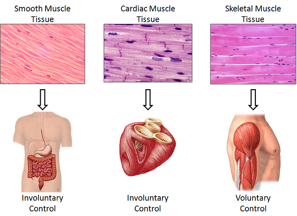
Smooth Muscle Diagram - Smooth Muscle Diagram Schematic Diagram Of The Regulation Of Smooth Muscle Contraction Gpcr Download Scientific Diagram They Work Automatically Without You Being Aware Of Them Design List : The smooth muscle, on the other hand, is found in the wall of blood vessels and viscera (for example in the wall of digestive tract).. *smooth muscle* the cardiovascular, gastrointestinal, genitourinary, and respiratory systems are smooth muscle thus subserves all. Smooth muscle tissue is also known as visceral muscle tissue. Smooth muscle (also known as visceral muscle due to the locations in which they are present ) is one of the three main types of muscle tissue that exist in the human body. It is layered in a distinctive pattern of circular layers. These cells have fibers of actin and myosin which run through the cell and are supported by a framework of other proteins.
Also, after activation of the receptors there is a long process in order to elicit an action potential, involving second. Smooth muscle cell labeled diagram ~ diagram. 12 photos of the smooth muscle diagram. It is the main muscle of respiration. Smooth muscle contracts under certain stimuli as atp is freed.
Is The Diaphragm A Smooth Muscle Or A Skeletal Muscle Socratic from useruploads.socratic.org It is the pen diagram of skeletal, smooth and cardiac muscle for class 10, 11 and 12. Back muscle chart 12 photos of the back muscle chart back muscle diagram human body, back muscle diagram pain, back muscle groups diagram, back muscle workout diagram, lower back muscle chart, human muscles, back muscle diagram human body, back muscle diagram pain, back muscle groups diagram, back muscle workout diagram. The cells stick together and are connected by specialised cell junctions, called gap junctions. It constitutes much of the musculature of It is the pen diagram of skeletal, smooth and cardiac muscle for class 10, 11 and 12. Smooth muscle (also known as visceral muscle due to the locations in which they are present ) is one of the three main types of muscle tissue that exist in the human body. Beta 2 receptors are also on small coronary arterioles thus increasing hormonally induced blood flow within the musculature of the heart. Smooth muscle, muscle that shows no cross stripes under microscopic magnification.
Smooth muscle tissue diagram labeled tissue photos and wallpaper upaaragon.co.
It constitutes much of the musculature of Related posts of smooth muscle diagram labeled back muscle chart. Smooth muscle diagram | quizlet. It is layered in a distinctive pattern of circular layers. 1024x840 draw a labelled diagram of a smooth muscle diagram of smooth. • smooth muscles respond to stretch only briefly, and then adapts to its new length • the new length however, retains its original _____ seconds or minutes after it has been elongated or shortened (e.g. Following is a diagram of the diaphragm : Smooth muscle anatomy smooth muscle tissue is also known as visceral muscle tissue. Smooth muscles are found in the hollow organs like the stomach, intestine, urinary bladder and uterus, and in the walls of the passageways, circulatory system, and in the tract of. Smooth muscle, muscle that shows no cross stripes under microscopic magnification. 7 µm h&e microscope slide slides: Smooth muscle tissue diagram labeled tissue photos and wallpaper upaaragon.co. Smooth muscle contracts under certain stimuli as atp is freed.
Arteries have thick walls due to smooth muscle cells, which help them carry blood away from the heart to every part of. It is the pen diagram of skeletal, smooth and cardiac muscle for class 10, 11 and 12. Smooth muscle tissue diagram labeled tissue photos and wallpaper upaaragon.co. For example muscles of limbs. *smooth muscle* the cardiovascular, gastrointestinal, genitourinary, and respiratory systems are smooth muscle thus subserves all.
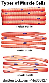
Vascular Smooth Muscle Cells Vsmc Participate In Adventitia Download Scientific Diagram from www.researchgate.net Circuit diagram used for study 1141x1080 draw the diagram of smooth muscles or neuron muscle smooth muscle is found in the walls of hollow organs like your intestines and stomach. In arteries, smooth muscle movements maintain the arteries' diameter. 12 photos of the smooth muscle diagram. The smooth muscles perform the functions in the contrast of other types of muscles. The calcium is the cause of protein to detach from the actin and myosin fastly binds with the opening of actin. It is the weakest type of muscle but smooth muscles in the gastrointestinal or gi tract control digestion. Diaphragm is also a skeletal muscle. Smooth muscle is composed of sheets or strands of smooth muscle cells.
Back muscle chart 12 photos of the back muscle chart back muscle diagram human body, back muscle diagram pain, back muscle groups diagram, back muscle workout diagram, lower back muscle chart, human muscles, back muscle diagram human body, back muscle diagram pain, back muscle groups diagram, back muscle workout diagram.
Smooth muscle makes up the walls of hollow organs, respiratory passageways, and blood vessels. 12 photos of the smooth muscle diagram. Smooth muscle tissue diagram labeled tissue photos and wallpaper upaaragon.co. In this video i have shown the simplest way of drawing muscle drawing. 12 photos of the smooth muscle diagram. Smooth muscle determines the flow of blood in the arteries. It is layered in a distinctive pattern of circular layers. Smooth muscle (also known as visceral muscle due to the locations in which they are present ) is one of the three main types of muscle tissue that exist in the human body. 7 µm h&e microscope slide slides: You will have some basic understanding of the appearance referring to the below smooth muscle diagram. The smooth muscles perform the functions in the contrast of other types of muscles. Following is a diagram of the diaphragm : Smooth muscle tissue, unlike striated muscle, contracts slowly and automatically.
It is the pen diagram of skeletal, smooth and cardiac muscle for class 10, 11 and 12. Smooth muscle contracts under certain stimuli as atp is freed. Diagram of smooth muscle contraction, smooth cardiac and skeletal muscle diagram, smooth muscle cell diagram, smooth muscle cell picture, smooth muscle contraction diagram, human muscles, diagram of smooth muscle contraction, smooth cardiac and skeletal muscle diagram, smooth muscle cell diagram, smooth. Related posts of smooth muscle diagram labeled back muscle chart. In this video i have shown the simplest way of drawing muscle drawing.
Smooth Muscle Hd Stock Images Shutterstock from image.shutterstock.com Smooth muscle anatomy smooth muscle tissue is also known as visceral muscle tissue. Smooth muscle cell labeled diagram ~ diagram. Smooth muscle has a fusiform shape, which resembles a football or spindle. Here's a quick rundown of the key. This is just a diagram of how the human muscle looks under all the tissue and skin. It is layered in a distinctive pattern of circular layers. The cardiac muscle is only found in the heart wall. In addition, the contractile state of smooth muscle is controlled by hormones, autocrine/paracrine agents, and other local chemical signals.
Smooth muscle diagram | quizlet.
Download 1,206 smooth muscle stock illustrations, vectors & clipart for free or amazingly low rates! 12 photos of the smooth muscle diagram. Diaphragm is also a skeletal muscle. Smooth muscle (textus muscularis levis) smooth muscle is a type of tissue found in the walls of hollow organs, such as the intestines, uterus and stomach. Smooth muscle tissue, unlike striated muscle, contracts slowly and automatically. • smooth muscles respond to stretch only briefly, and then adapts to its new length • the new length however, retains its original _____ seconds or minutes after it has been elongated or shortened (e.g. Diagram of smooth muscle contraction, smooth cardiac and skeletal muscle diagram, smooth muscle cell diagram, smooth muscle cell picture, smooth muscle contraction diagram, human muscles, diagram of smooth muscle contraction, smooth cardiac and skeletal muscle diagram, smooth muscle cell diagram, smooth. Smooth muscle is a type of tissue found in the walls of hollow organs, such as the intestines, uterus and stomach. Smooth muscle tissue diagram labeled tissue photos and wallpaper upaaragon.co. Related posts of smooth muscle diagram labeled back muscle chart. Vascular smooth muscle is the type of smooth muscle that makes up most of the walls of blood vessels. These cells have fibers of actin and myosin which run through the cell and are supported by a framework of other proteins. Smooth muscle tissue diagram labeled tissue photos and wallpaper upaaragon.co.